Burns & complex wounds
Tier-1 and 2 hospitals with active burns and wound units. Predictive grafts where tissue composition, bed readiness, and print params decide outcomes.
Now booking hospital and lab pilots. Work with us

Bioprint the exact graft your body needs. From a wound image to tissue mix, bioink, and print params - computed, not guessed.
Backed by researchers and early capital.
Partnered with research and clinical labs.
 Tvashtr BiotechTvashtr BiotechTvashtr Biotech
Generic grafts fail to integrate; autografts wound the donor site. Zyogen turns a photo into printable bioink, scaffold, and params. On-prem, DPDP & HIPAA ready.
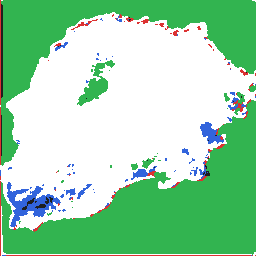
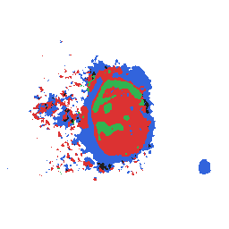
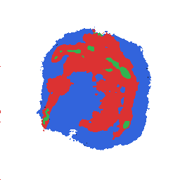
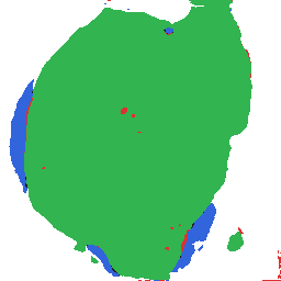
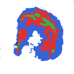
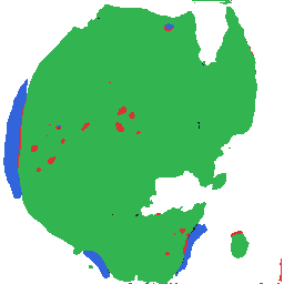
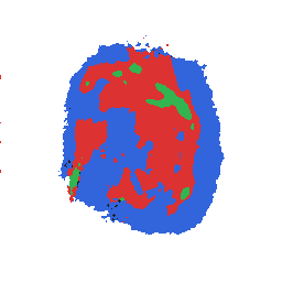
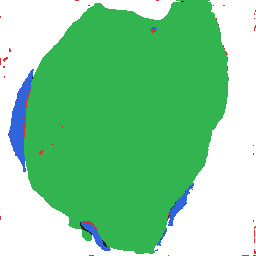
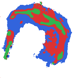
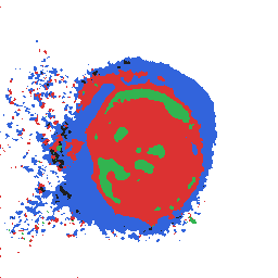
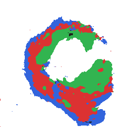
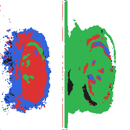
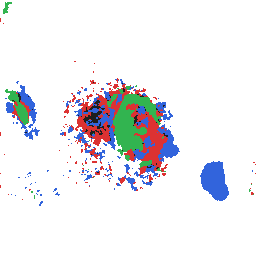
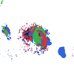
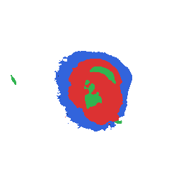
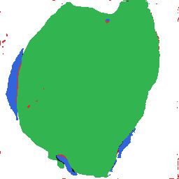
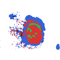
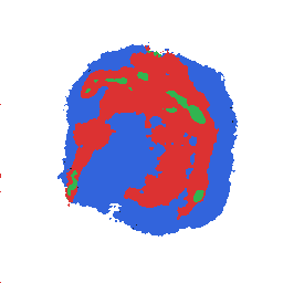
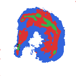
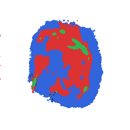
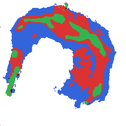
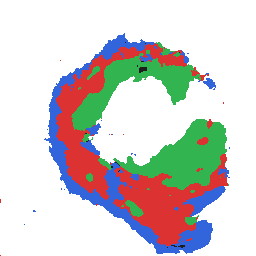
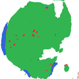
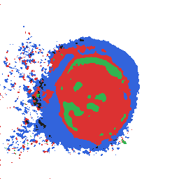
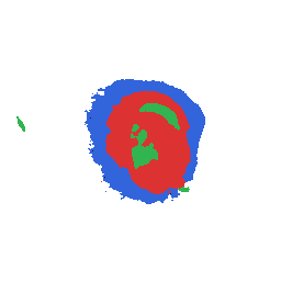
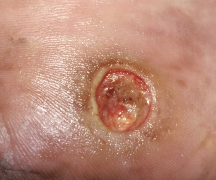
ViT locked geometry and class patches. Majority granulation with fibrin islands and a thin epithelial rim.
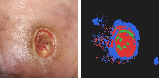
Area fractions set the bioink blend; physics scored the recipe inside the wet-lab envelope.
Area fractions drive bioink blend and scaffold porosity.
Recipe confidence and wound area after vision normalization.
Median recovery ~24 days. Scrub the timeline.
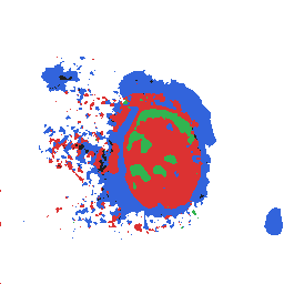
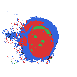
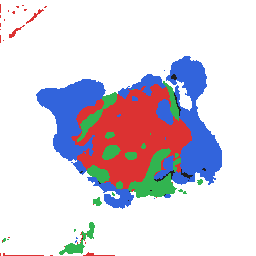
Diffusion conditioned on tissue mix and heal rate. Median recovery ~24 days - scrub the timeline.
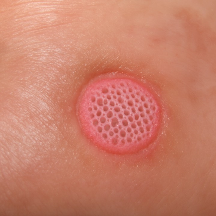
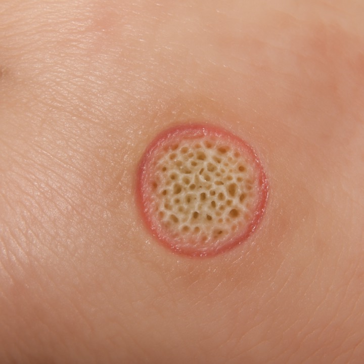
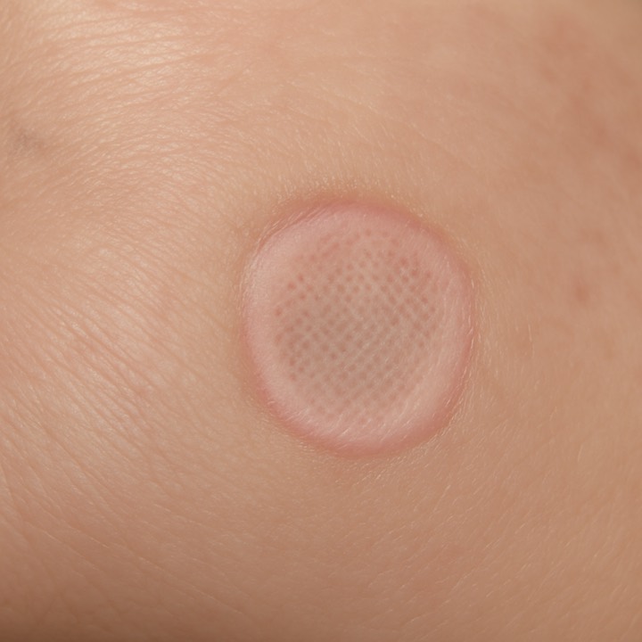
Curves are conditioned on day-0 tissue fractions and heal rate - they move with the timeline scrub, not measured follow-up imaging.
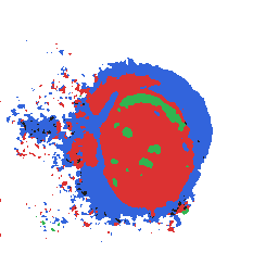
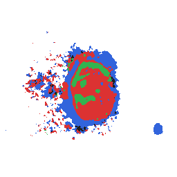
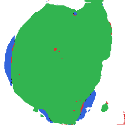
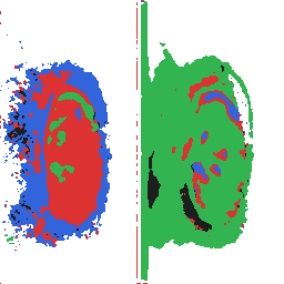
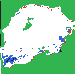
A vision model maps the bed patch by patch: granulation, fibrin, epithelium, necrosis. Graft design starts from what is actually there.
We audit model internals so predictions key on real tissue biology, across the tissues we print, not rulers, lighting artifacts, or demographic shortcuts.
Tier-1 and 2 hospitals with active burns and wound units. Predictive grafts where tissue composition, bed readiness, and print params decide outcomes.
Corneal and ocular surface programs adopting clinical imaging workflows. Clear SaMD-style routes for prediction software beside the surgeon.
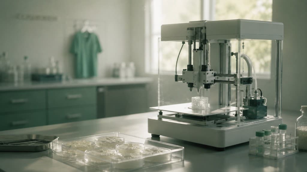
Cartilage and soft-tissue reconstruction teams already running bioprinters from Cellink, Regemat, 3D Systems, and NBIL in research hospitals.
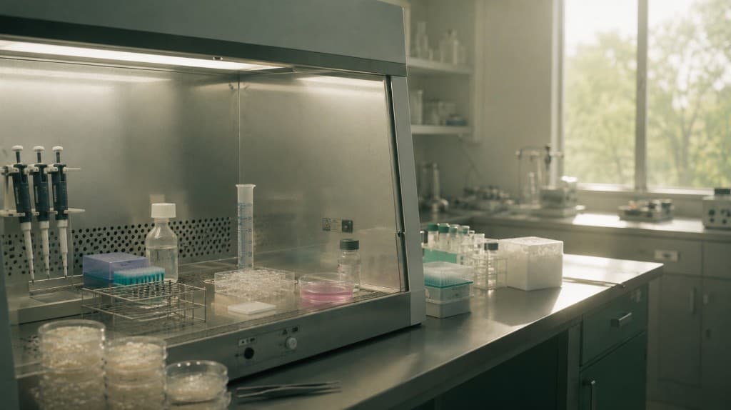
Tissue engineering labs at IITs and AIIMS. India R&D runs 70 to 80% cheaper: build the stack here, then export the method.

Secondary expansion markets where India’s cost advantage compounds. Same hospital SaaS + per-graft model, priced below imported stacks.
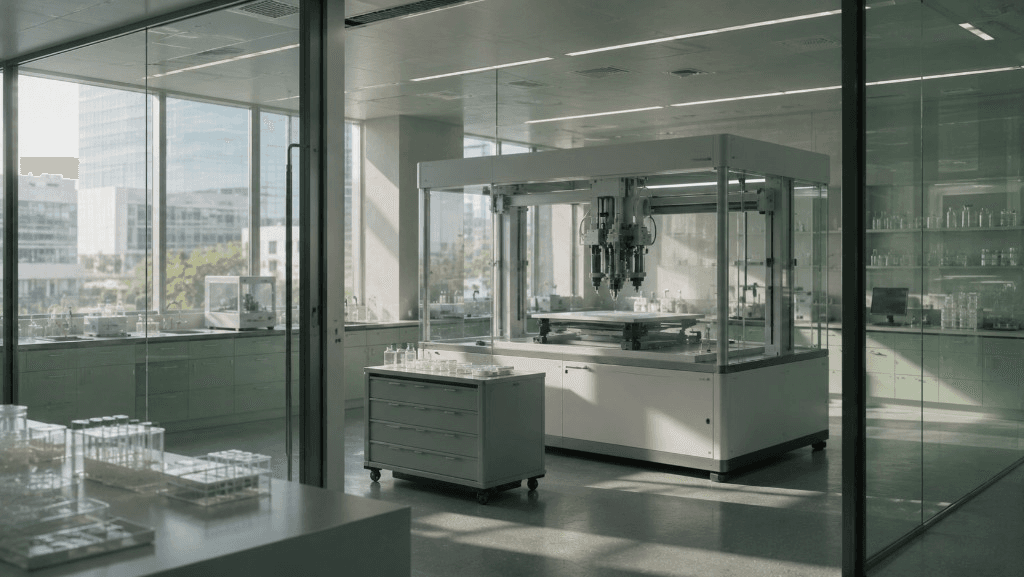
Global expansion after proving deployment in India. FDA SaMD and EU MDR routes for clinical prediction software already exist.
product and tissue engineering.
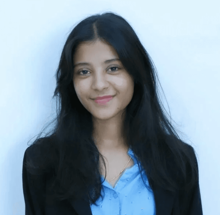
Founder & CEO
Built and exited ImpactPlay for cancer patients. Robotics and medtech projects with acquired IP. AI Growth & Insights @ Google (via Canvas8). IEO International Rank 6. 3× valedictorian.
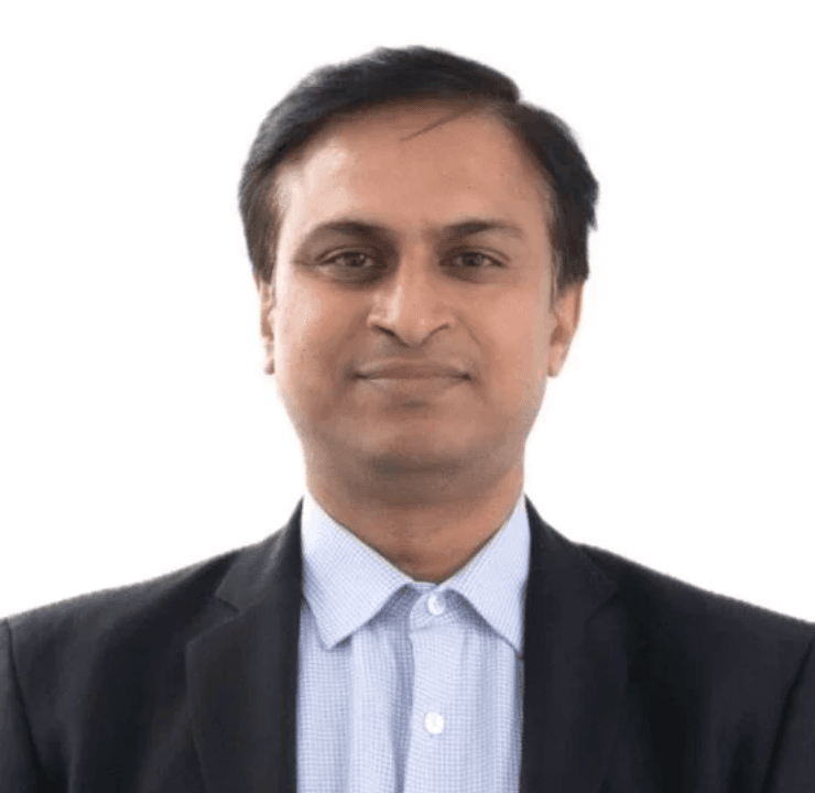
Prof. Falguni Pati
Scientific Collaborator
HOD Biomedical Engineering, IIT Hyderabad. 7,700+ citations, 100+ publications. First author, Nature Communications (2014) on decellularized-ECM bioinks.
Learn more about predictive bioprinting with Zyogen.
No. Zyogen automates structure, consistency, and parameter suggestion. Your clinical team leads interpretation and the final graft decision.
No. The stack is deterministic. A physics engine computes bioink formulation, scaffold geometry, and print parameters from wet-lab data, not hallucinated outputs.
A vision model maps wound-bed composition, granulation, fibrin, necrosis, and related conditions. Those fractions feed predictors for whether the bed can take a skin graft and how healing is likely to run.
Medical models can secretly key on rulers, skin tone, or lighting. We audit internals so the system is trusted for the right biological reasons before it informs a decision.
Sovereign deployment: self-hosted and on-prem inside hospital infrastructure, designed for DPDP and HIPAA compliance.
Yes. A hardware translation layer produces toolpaths and extrusion protocols for multi-modal bioprinters already installed in research and clinical settings.
Hospitals, research labs, and partners building the predictive layer for regenerative care.